
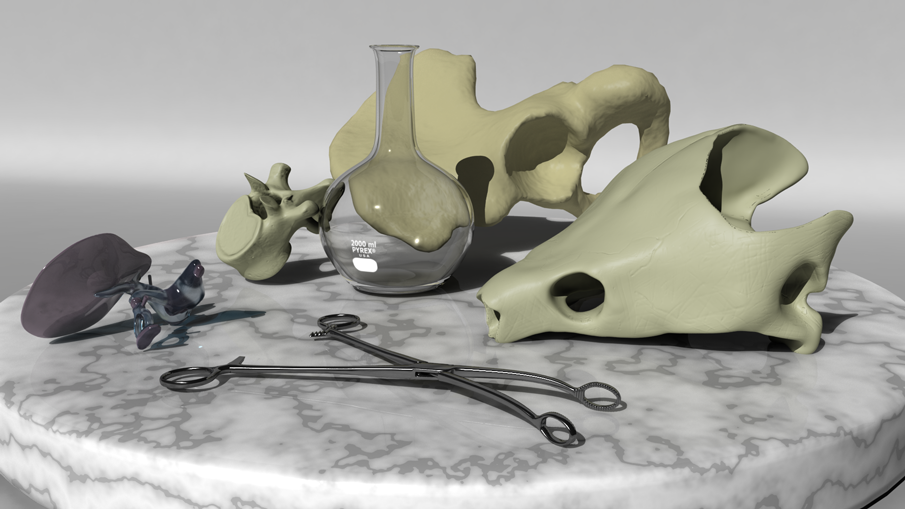
Hover over the individual pieces in the image below for identification.
This scene was organically modeled in Maya. The lighting was generated using a high dynamic range (HDR) image.
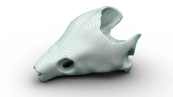
1. Turtle skull: An actual turtle skull (unknown species) was scanned using a 3D scanner and retopologized in 3D Coat. Some texture was added in Mudbox.
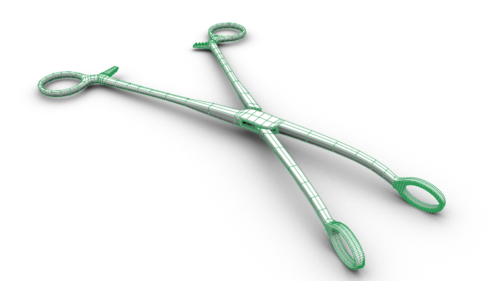
2. Sponge forceps: These were modeled organically in Maya as a polygon object and the actual instrument for reference.
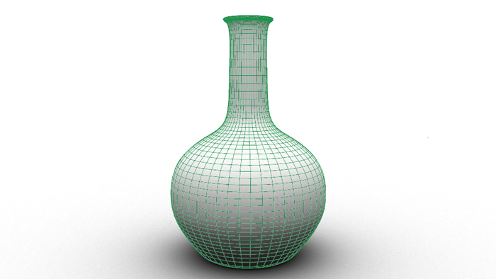
3. Flask: This was modeled in Maya using NURBs, with an actual flask as reference.
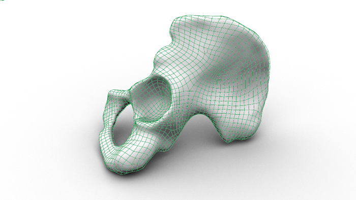
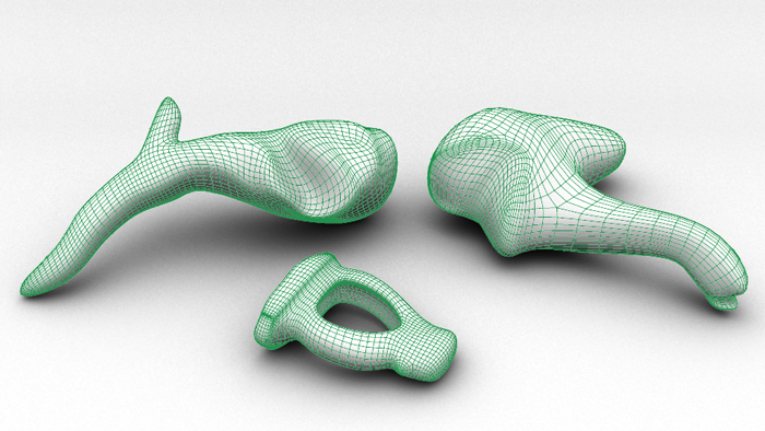
4. Pelvic Bone: A reconstruction of a CT scan was was made in Osirix originally. Then the reconstruction was imported into 3D Coat to be retopologized, then textured in Maya.
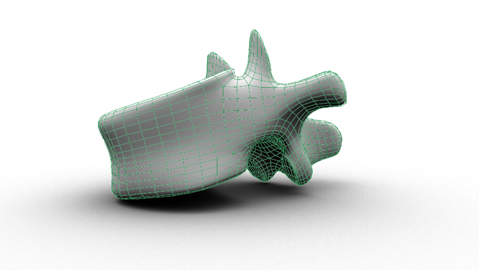
5. Vertebra: This was modeled organically in Maya using polygons and an actual vertebra as a reference. Some texturing was done in Mudbox.
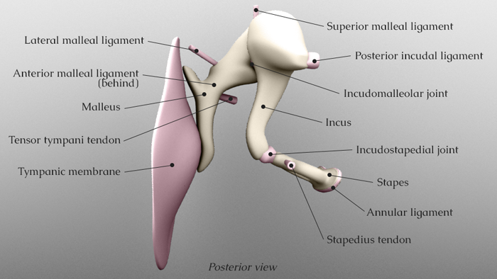
6. Middle Ear: All components of the middle ear (malleus, incus, stapes, tympanic membrane, incudostapedial joint, incudomalleolar joint, tensor tympani tendon, stapedius muscle tendon, annular ligament, posterior incudal ligament, lateral malleal ligament, superior malleal ligament, and anterior malleal ligament) were modeled organically using polygons from references listed below.
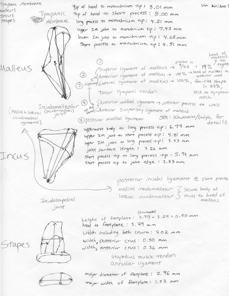
1. Dass, R., Grewal, B. S., & Thapar, S. P. (1966). Human stapes and its variations. I. General features. Journal of Laryngology Otology, 80, 11-25.
2. Dass, R., Grewal, B. S., & Thapar, S. P. (1966). Human stapes and its variations. II. Footplate. The Journal of laryngology and otology, 80(5), 471.
3. Funnell, W. R. J. (2013, August 16). Auditory Mechanics Laboratory. Retrieved October 21, 2013, from http://audilab.bmed.mcgill.ca/AudiLab/
4. Graboyes, E. M., Hullar, T. E., & Chole, R. A. (2011). The lenticular process of the incus. Otology & neurotology: official publication of the American Otological Society, American Neurotology Society [and] European Academy of Otology and Neurotology, 32(9), 1600.
5. Gulya, A. J. (2007). Gulya and Schuknecht’s anatomy of the temporal bone with surgical implications. Informa healthcare.
6. Kirikae, I. (1960). The structure and function of the middle ear. Tokyo: University of Tokyo Press.
7. Weistenhöfer, C., & Hudde, H. (1999). Determination of the shape and inertia properties of the human auditory ossicles. Audiology and Neurotology, 4(3-4), 192-196.